ALERT
ALERT Attention UNM Users: On March 9th, UNM iLab URL changed to unm.ilab.agilent.com where you will sign in using UNM or HSC credentials. Please bookmark new URL.
The Banner financial integration went live on March 9th which includes transitioning from PR Numbers to Indexes.
Please contact iLab Support (ilabsupport@agilent.com) for assistance and questions.
Restricted and Unrestricted Indices (non grant funded) can now be requested into a PI's lab.
Note: For Institutional Administrators:
Please DO NOT accept lab requests unless you are assisting a PI. Thank you
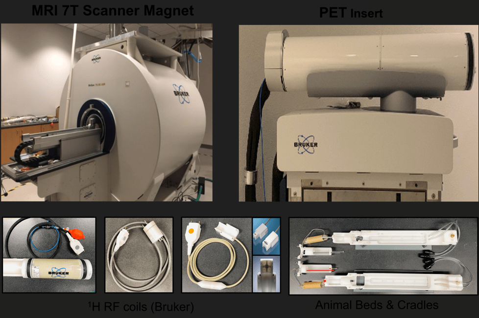
(hide this warning on this page)The Preclinical Imaging Core at Domenici Hall houses a 7T MRI scanner (Bruker BioSpec 70/30USR) with an integrated PET insert for preclinical and molecular in vivo and ex vivo imaging. The MRI scanner is equipped with state-of-the-art multi-channel RF coils, allowing high-resolution imaging for applications in life sciences, biomedical research, and preclinical studies. The PET system enables highly sensitive detection of radiotracers, supporting functional and molecular studies in small animal models. The simultaneous acquisition of PET and MR images combines the high sensitivity of PET with the high spatial resolution and soft tissue contrast of MRI. The facility provides comprehensive imaging services for both UNM and external investigators.
Our 7T MRI/PET system includes a complete set of three efficient gradient coils. Task designated imaging RF coils (Volume and Surface) are available to improve the MR image quality. For simultaneous PET and MR imaging, the PET module is inserted within the bore of the MRI system for automatic imaging from one modality to another on the same axis. An isofluorane anesthesia system, body temperature control, and vital signs monitoring system (i.e., temperature, respiration and ECG) plus an MRI-compatible small animal gating system (SA Instruments) are installed to permit free-breathing acquisition during quantitative MRI measurements.
Core services offered:
1. Imaging at 7 Tesla Bruker PET/ MRI
• In-vivo and ex-vivo MR imaging studies in rodents and other small laboratory animals
• 1H Magnetic Resonance Imaging, 1H Magnetic Resonance Spectroscopy
• FDG- PET imaging
2. Study initiation processes
• Discuss the research idea and goals
• Core users work with imaging scientists to develop a detailed study design
• Support for animal protocol submissions to IACUC
• Experimental set-up and feasibility testing and Optimization of imaging protocols
3. Data acquisition and analysis
• Customized to achieve study goals
• Assist with imaging data interpretation
4. Consultation and support for imaging grant applications
5. Support for manuscript preparation
6. Training for faculty and students in the imaging method
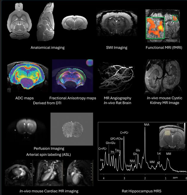
MRI Imaging:
• High-resolution structural and functional imaging in rodents and ex vivo tissues
• Anatomical imaging: T1, T2, T2*, and proton density mapping
• Diffusion MRI: DWI and DTI for tissue microstructure and fiber tracking
• Functional MRI: BOLD and arterial spin labeling (ASL) for perfusion and activation mapping
• Non-invasive Perfusion Imaging: ASL, CASL
• Angiography and susceptibility-weighted imaging (SWI)
• Magnetic Resonance Spectroscopy (MRS): In vivo metabolic profiling
Applications include models of stroke, TBI, vascular dementia, Alzheimer’s disease, Normal development and aging, Detection/Monitoring of lesions (tumor, ischemia, etc.), Body fat distribution, Cerebral blood flow, Magnetic resonance angiography (MRA), Evaluation of brain glucose metabolism in neurodegenerative disorders etc.
PET/MRI Imaging:
• Three-ring PET Insert (Si198) for Simultaneous PET/MR
• In vivo molecular imaging using [18F]-FDG and other radiotracers
• Evaluate tumor metabolism, neurodegeneration, and organ function
• Integrated acquisition for co-registered PET/MR datasets
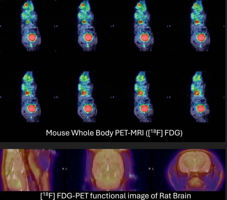
Image Processing:
Two dedicated workstations are available for data transfer and offline data processing. Commercial and open-source software are available includes
• Bruker’s PV 7.0, PV360 v3.5 and PMOD
• MATLAB (MathWorks Inc.)
• ImageJ (NIH, Bethesda, MD)
• SPM, FSL, etc.
Munish Chauhan, PhD
Director, Preclinical Imaging Core (MRI/PET)
Research Assistant Professor, Department of Radiology
mchauhan@salud.unm.edu
480-799-6611
| Hours | Location |
|
Monday - Friday 8am - 5pm |
Pete and Nancy Domenici Hall 1101 Yale Blvd NE Albuquerque, NM 87106-0001 |
For more information, Click Here.
| Name | Role | Phone | Location | |
|---|---|---|---|---|
| Munish Chauhan |
Director
|
505-272-9919
|
mchauhan@salud.unm.edu
|
Pete and Nancy Domenici Hall Room Numbers: 1121-1123 1101 Yale Blvd NE Albuquerque, NM 87106-0001
|